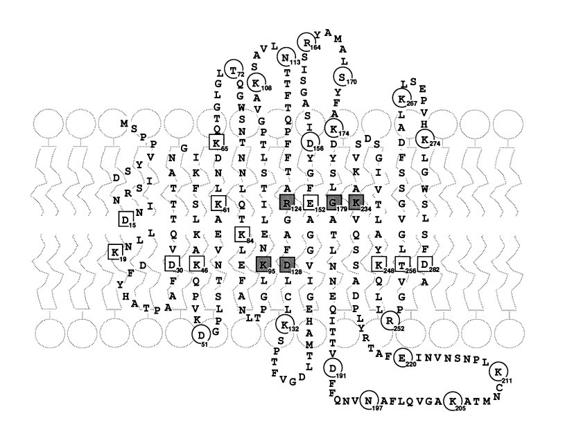
 Fig. 8. Folding pattern
of yeast VDAC in a phospholipid membrane. This pattern illustrates the
transmembrane strands proposed to form the walls of the pore of the
VDAC channel. The extended b
strands would be hydrogen-bonded together and to the a
helix forming a cylindrical structure. The boxed residues are sites
that
affect the ion selectivity of the channel. Circled
residues are sites that had no effect on ion selectivity.
Fig. 8. Folding pattern
of yeast VDAC in a phospholipid membrane. This pattern illustrates the
transmembrane strands proposed to form the walls of the pore of the
VDAC channel. The extended b
strands would be hydrogen-bonded together and to the a
helix forming a cylindrical structure. The boxed residues are sites
that
affect the ion selectivity of the channel. Circled
residues are sites that had no effect on ion selectivity.