Welcome to the Jeffery Lab!The Jeffery lab studies (1) the molecular and genetic basis of evolutionary change in development and (2) the mechanisms of regeneration. |
|
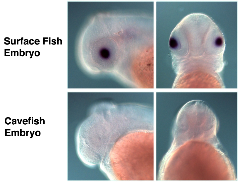
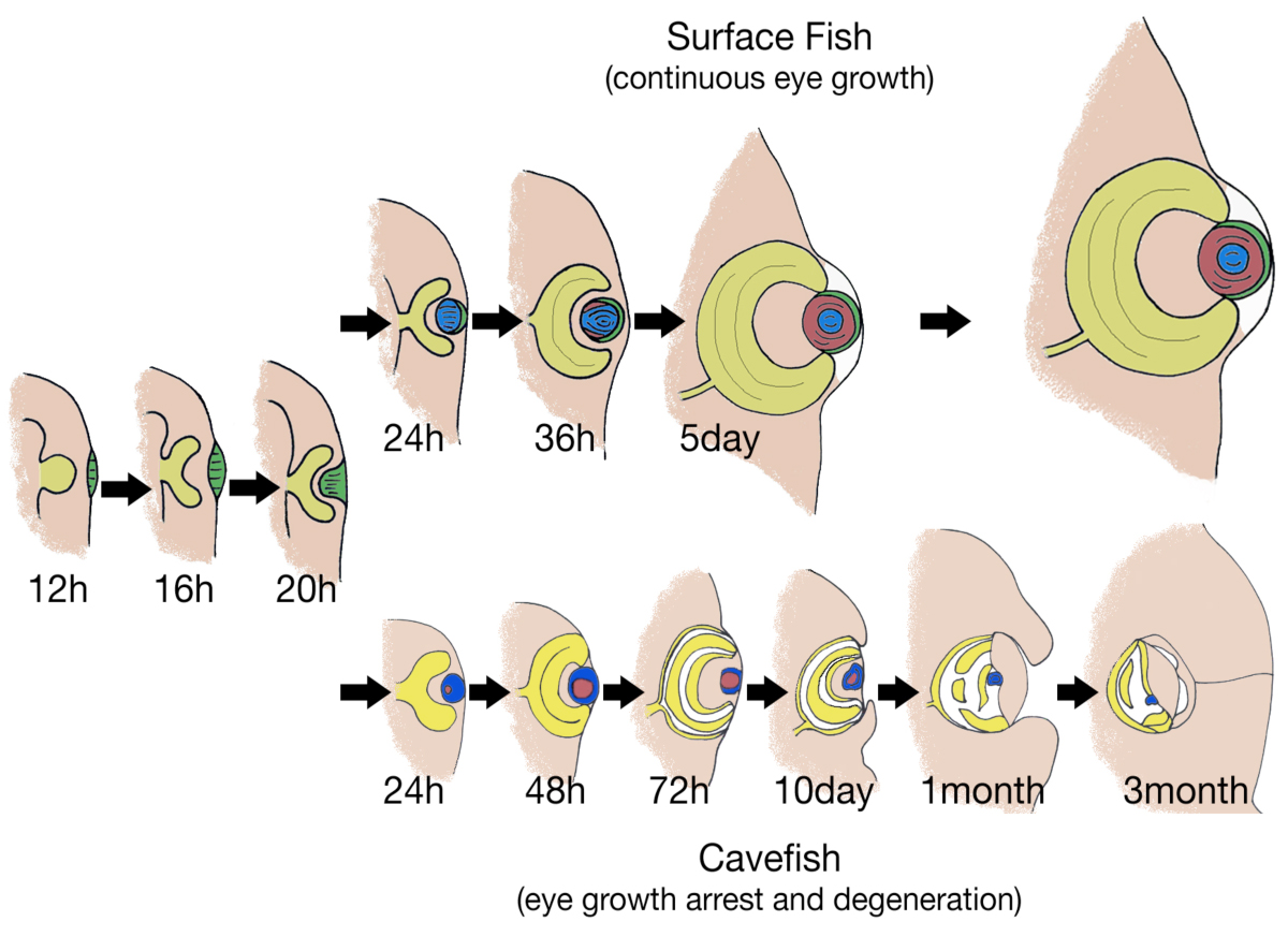
Evolution of Development We are interested in determining how and why cavefish have lost their eyes and pigmentation. By genetic and
genome approaches, we are analyzing genes associated with quantitative trait loci (QTL) with the objective of finding mutations involved in eye loss, which is a complex genetic trait controlled by about a dozen gene loci. Using this information we address the evolutionary forces involved in eye degeneration. Although many mechanisms may be involved, thus far we have evidence that eye loss is antagonistically linked as a pleiotropic tradeoff with constructive traits, such as taste buds and sensory neuromasts, which are adaptive in the cave environment. |
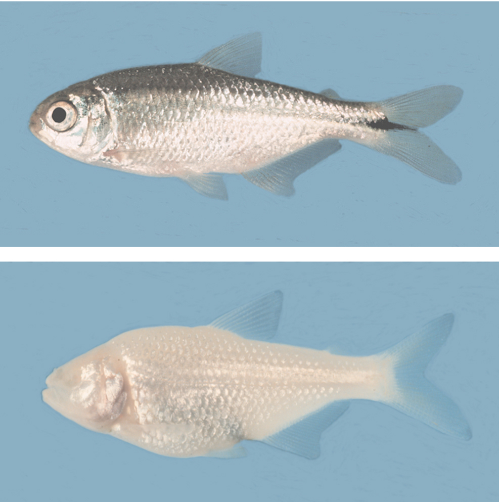
The surface dwelling and cave dwelling forms of Astyanax mexicanus. The cave form has lost eyes and melanin pigmentation. For details see any of our Astyanax publications. |
 |
|
|
|
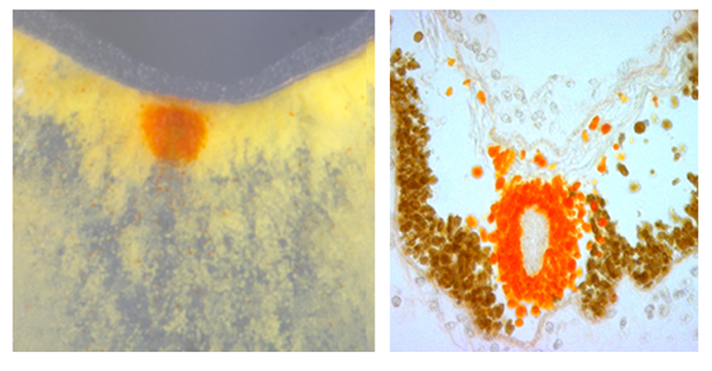
Expression of the anti-apoptotic chaperone aA-crystallin is downregulated in the cavefish lens. This is thought to be one of the reasons for lens apoptosis and eye degeneration in cavefish. See Ma et al. (2014) for details.
|
We are also studying the parallel/convergent evolution of eye and pigment regression in different Astyanax cavefish populations that have evolved these traits independently. We have obtained evidence that the loss of melanin pigmentation (albinism) in cavefish is regulated at the first step of melanin synthesis by mutations in the oca2 gene. One of the benefits of blocking melanin synthesis at the oca2 step may be to shunt excess L-tyrosine into the alternative catecholamine synthesis pathway, which is enhanced in albino cavefish. |
| Regenerative Biology Using ascidians (tunicates or sea squirts), which are thought to be the sister group of vertebrates, we study the molecular and cellular basis of tissue repair and regeneration. These studies are carried out on the model ascidian Ciona intestinalis, which has a rich of history of research in developmental biology, a sequenced genome, and offers many attractive molecular and genomic tools for studies on regeneration. We have found that distal structures of the adult animal, namely the siphons and brain, regenerate rapidly and with a great deal of fidelity after injury or removal. |
|
|
|
Distal regeneration requires the participation of stem cells located in the underlying branchial sac (pharynx), which are induced to proliferate as response to injury, producing progenitor cells that migrate and form a blastema at the wound site. The blastema cells later differentiate into distal tissues, including musculature, nerve cells, and sensory organs. Recent studies have shown that the Notch signalling system is involved in this process. We have also demonstrated that the capacity to regenerate, which is robust in young adults, gradually fades and finally disappears in old animals. Thus, the Ciona system is also under development as model to address the mechanisms underlying the loss of tissue replacement and regenerative potential during aging. The latter studies are being carried out in a satellite laboratory in the Bell Center of Regenerative Biology and Tissue Engineering at the Marine Biological Laboratory in Woods Hole, MA.
[Home] - [Contact Us] - [Web Accessbility] |
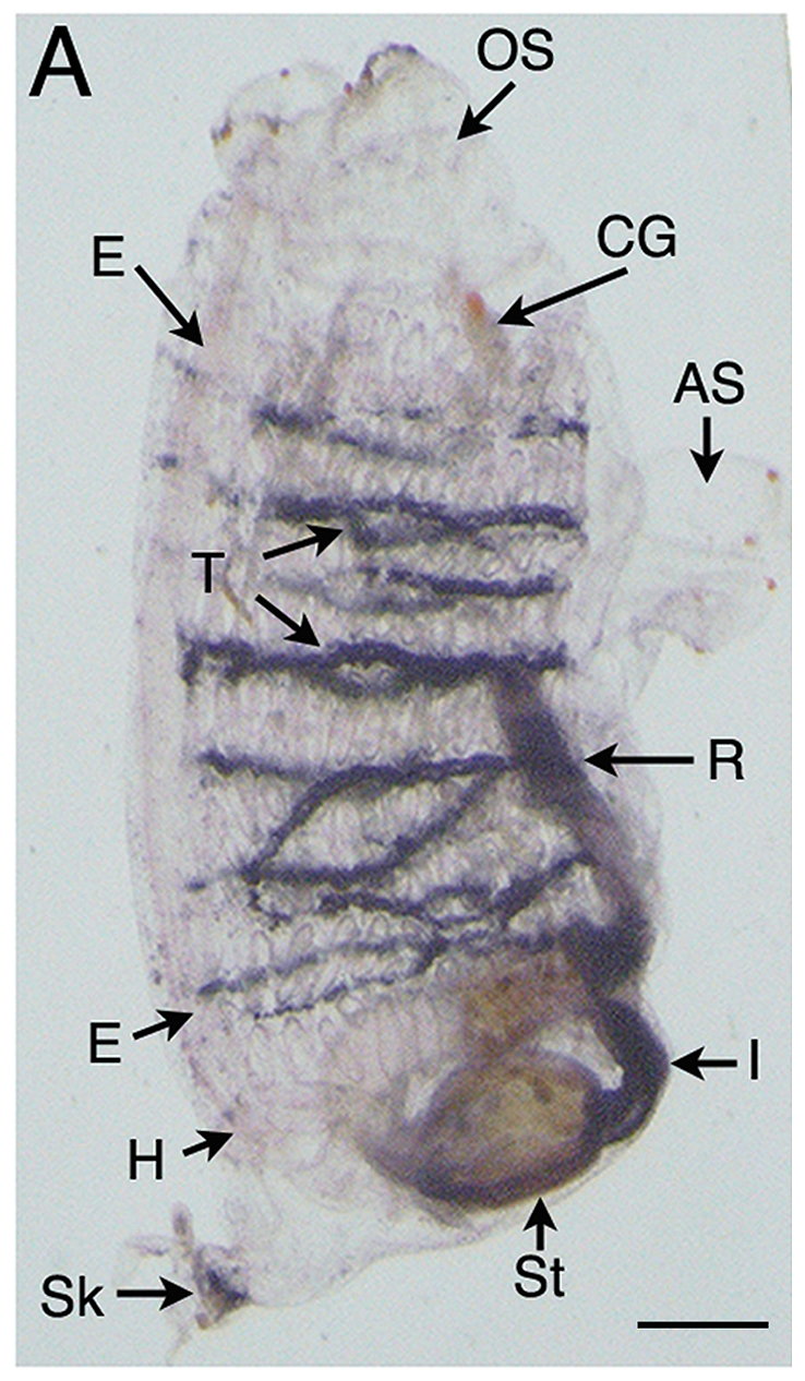
An adult Ciona showing blue stained stem cell niches in transverse vessels (T) of the branchial sac. These stem cells are involved in replacement of the oral siphon during regeneration. See Jeffery (2014) for details. |
![]()