Lab 6. Agglutination

            When antibodies are mixed with their corresponding antigens on the surface of large, easily sedimented particles such as animal cells, erythrocytes, or bacteria, the antibodies cross-link the particles, forming visible clumps.  This reaction is termed agglutination.  Agglutination is a serological reaction and is very similar to the precipitation reaction we learnt last week.  Both reactions are highly specific because they depend on the specific antibody and antigen pair.  The main difference between these two reactions is the size of antigens.  For precipitation, antigens are soluble molecules, and for agglutination, antigens are large, easily sedimented particles.  As you will see from this lab exercise, agglutination is more sensitive than precipitation reaction because it takes a lot of more soluble antigens and antibody molecules to form a visible precipitation.  To make the detection of soluble antigen and antibody reaction more sensitive, a precipitation reaction can be transformed into an agglutination reaction by attaching soluble antigens to large, inert carriers, such as erythrocytes or latex beads.

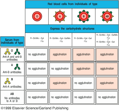
            Agglutination reactions have many applications in clinical medicine.  Agglutination reactions can be used to type blood cells for transfusion, to identify bacterial cultures, and to detect the presence and relative amount of specific antibody in a patient’s serum.  Agglutination has been commonly used to determine whether a patient had or has a bacterial infection.  For example, if a patient is suspected of having typhoid fever, the patient’s serum is mixed with a culture of Salmonella typhi.  If an agglutination reaction occurs, shown as clumping of the bacteria, the patient either had or has an S. typhi infection.  Since certain antibodies can persist in a patient’s blood for years after the patent has recovered from the infection, a positive reaction does not mean that the patient currently has the infection.  To determine whether a patient is currently suffering from typhoid fever, the amount or titer of the antibody will be determined at the onset of illness and two weeks later.  If the titer of antibody in the patient’s serum has increased at least four-fold between the two tests, the patient is currently fighting off the infection, and the pathogen causing the illness is confirmed.

             In this lab exercise, you will learn two different methods of employing agglutination reactions, rapid slide agglutination and microtiter test.  These two tests are valuable methods commonly used in clinical laboratories.  Applications of agglutination include A-B-O blood typing tests and rapid bacterial identification.  The microtiter test is used to quantify the amount of antibody in patient’s blood.


Part 1.     Rapid Slide Agglutination

          In this lab exercise, you will learn how to use rapid slide agglutination to determine your blood type.  The surface of blood cells of each type expresses a unique oligosaccharide structure that is called A or B antigens.  The A and B antigen are inherited.  Each person carries anti-A and/or anti-B antigen in his or her blood dependent on his or her blood type.  Presence in the blood of anti-A and/or anti-B antibodies is not inherited, but is the result of (1) prior exposure to A- and B-like antigens in the environment, such as bacterial surface molecules; or (2) an immunological response to nonself molecules.  When a person receives unmatched blood, these antibodies will either agglutinate blood cells or induce complement-mediated cytolysis.  Typing blood to match donor and recipient with respect to ABO antigens is an important and widely used procedure.
 
        SAFETY NOTE: This lab uses your own blood for ABO blood typing.  After obtaining your drops of blood, put on gloves.  Dispose of waste in the biohazard container.

Procedure

1. Label three slides A, B and A+B.

2. Spray your left “ring “ finger with 70% ethanol or wipe it with an alcohol wiper and let it air dry.

3. Take a sterile lancet and puncture your fingertip.  If you have calluses, aim a little to the side.  DO NOT LANCET ANYONE OTHER THAN YOURSELF.  When finished with your lances, place them in the BIOHAZARD CONTAINER.

4. Place a small drop of blood on each of three microscope slides.  At this point, put on GLOVES.  DO NOT TOUCH ANYONE ELSE’S BLOOD.

5. On the A slide, place a 20 ml of anti-A antiserum.  Place anti-B on the B slide and anti-A + anti-B on the A+B slide. Mix the antisera in with the blood using a separate toothpick for each slide.  Place toothpicks in the biohazard waste.

6. After several minutes, observe agglutination and determine your blood type.


Part 2.     Microtiter Test

            In this lab exercise, you will learn how to use the microtiter test to determine the amount of anti-sheep red blood cell (RBC) antibody.

Procedure

1. In a round-bottom microtiter plate, add 50 ml of PBS to columns 2-9 in rows A-C.  Leave column 1 empty.

2. Add 100 ml of rabbit anti-sheep RBC to column 1 in rows A-C.

3. In each row, transfer 50 ml from column 1 to column 2 and mix well.   Now take 50 ml from column 2 and transfer to column 3.  Repeat this process across the columns.  Discard the final 50 ml taken from row 9.

4. Add 50 ml of a 2% suspension of sheep RBC to all wells (columns 1-9, rows A-C). Make sure the RBCs stay adequately resuspended as you are using them.  Periodically invert the capped tube to keep them resuspended evenly.

5. Incubate the plate for 24 hours at room temperature.

6. Come back the next day and observe agglutination.  Positive wells will exhibit a diffuse and confluent settling of the RBC and Ab, while in negative wells, all cells will roll down to the bottom and it will look more like a dot.
 

Reading
Immunobiology A-7

Study Questions

1.  What is the difference between precipitation and agglutination?  Which one of these two methods is more sensitive and why?

2.  What type of structure on your red blood cells decides your blood type?
 

3.  You used the slide agglutination test to determine your blood type.  You got the following result:
     Anti-serum      A     B     AB
     Agglutination    -     +       +

 What is your blood type?

If you receive type AB blood for blood transfusion, what is going to happen and why?

4. Describe how to determine whether a patient has ever had a Salmonella typhi infection, the name of method, reagents used, possible results, conclusions deduced from results and why.

5. In preparing her immunology lab, an instructor purified IgG antibodies that are specific to sheep red blood cells (SRBCs) and digested some of the antibodies into Fab, Fc, and F(ab’)2 fragments.  She placed each preparation in a separate tube, labeled the tubes with a water-soluble marker, and left them in an ice bucket.  When the instructor returned, she discovered that the labels had smeared and were unreadable.  Determined to salvage the antibodies, she relabeled the tubes 1, 2, 3, and 4 and proceeded.  Based on the test results described below, indicate which preparation was contained in each tube and explain why you so identified the contents.

a. The preparation in tube 1 agglutinated SRBCs but did not lyse them in the presence of complement.

b. The preparation in tube 2 did not agglutinate SRBCs or lyse them in the presence of complement.  However, when this preparation was added to SRBCs before the addition of whole anti-SRBC, it prevented agglutination of the cells by the whole anti-SRBC antiserum.

c. The preparation in tube 3 agglutinated SRBCs and also lysed the cells in the presence of complement.

d. The preparation in tube 4 did not agglutinate or lyse SRBCs and did not inhibit agglutination of SRBCs by whole anti-SRBC antiserum.